
More Information
Submitted: June 25, 2025 | Approved: July 04, 2025 | Published: July 07, 2025
How to cite this article: Tlili Y, Mabrouk A, Mroua B, Dhaw AB, Kbir GH, Moussa MB. Giant Adult-onset Juvenile Xanthogranuloma in an Unusual Location Arch Surg Clin Res. 2025; 9(2): 025-026. Available from:
https://dx.doi.org/10.29328/journal.ascr.1001087.
DOI: 10.29328/journal.ascr.1001087
Copyright license: © 2025 Tlili Y, et al. This is an open access article distributed under the Creative Commons Attribution License, which permits unrestricted use, distribution, and reproduction in any medium, provided the original work is properly cited.
Keywords: Giant; Juvenile xanthogranuloma; Adult; Histiocytosis; Excision

Giant Adult-onset Juvenile Xanthogranuloma in an Unusual Location
Yassine Tlili1*, Aymen Mabrouk1, Bassem Mroua2, Anis Ben Dhaw1, Ghassen Hamdi Kbir1 and Mounir Ben Moussa1
1Department of General Surgery A21, Charles Nicolle Hospital, Faculty of Medicine of Tunis, Tunisia
2Department of General Surgery, Menzel Tmim Hospital - Nabeul, Faculty of Medicine of Tunis, Tunisia
*Address for Correspondence: Yassine Tlili, Department of General Surgery A21, Charles Nicolle Hospital, Faculty of Medicine of Tunis, Tunisia, Email: [email protected]
We report the case of a 29-year-old male referred to our surgical department for evaluation of two progressively enlarging lumbar masses with an eight-month history. Physical examination revealed two firm, non-tender subcutaneous nodules in the bilateral lumbar regions (Figure 1), measuring 9 cm and 8 cm in their greatest dimensions, respectively, with deep adherence to the underlying muscular fascia. Magnetic Resonance Imaging (MRI) identified two irregular, well-circumscribed soft tissue masses displaying T2-weighted hyperintensity and homogeneous contrast enhancement following Gadolinium administration, both intimately associated with the adjacent musculature (Figure 2). Due to the substantial lesion size and resultant patient discomfort, surgical excision was performed with a margin of surrounding muscular tissue to ensure complete resection (Figure 3). The postoperative course was unremarkable. Histopathological examination demonstrated a polymorphous inflammatory infiltrate predominantly composed of histiocytes with occasional multinucleated giant cells, confirming the diagnosis of Juvenile Xanthogranuloma (JXG). No recurrence was observed during twelve months of postoperative follow-up.
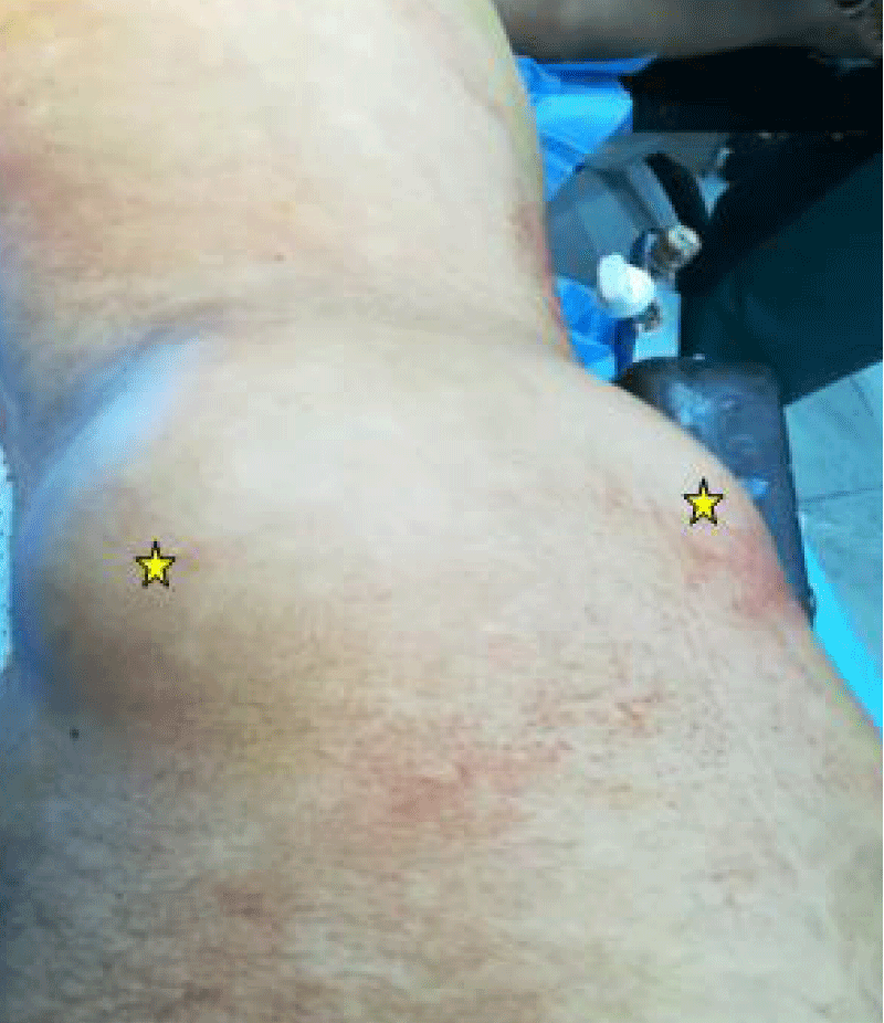
Figure 1: Clinical examination revealing two lumbar masses (*).
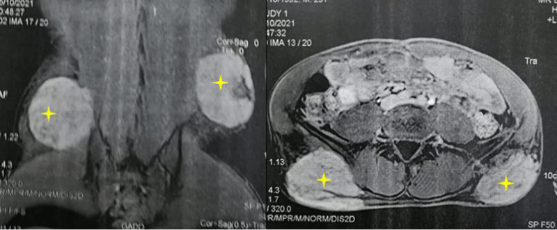
Figure 2: MRI demonstrating two lumbar soft tissue masses with T2 hyperintensity and Gadolinium enhancement (+).
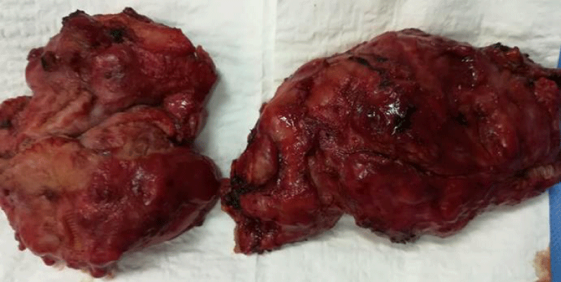
Figure 3: Resected specimens.
Juvenile xanthogranuloma is a benign, non-Langerhans cell histiocytosis, typically appearing as self-limiting cutaneous papules or nodules in infants, with most cases arising within the first year of life [1,2]. While the head and neck are the most commonly affected sites, lesions may also appear on the trunk, extremities, or, rarely, in extracutaneous locations [2]. Histopathological examination reveals a characteristic infiltrate of histiocytes, foamy cells, and Touton giant cells. Although the etiology remains unclear, JXG is thought to represent a reactive histiocytic proliferation triggered by unknown stimuli [1].
This case highlights the diagnostic and therapeutic challenges of giant adult-onset JXG in an atypical location. Although JXG is typically a pediatric condition, our report underscores its potential occurrence in adults, necessitating wide surgical excision due to size and local invasiveness. Histopathology remains the gold standard for diagnosis. Long-term follow-up is essential to monitor recurrence, though our patient showed no signs after 12 months. Further studies are needed to elucidate the pathophysiology of adult-onset JXG.
Ethical declarations
Informed consent: Written informed consent was obtained from the patient for publication of this case and accompanying images.
We thank the patient for consenting to share this case. We also acknowledge the contributions of the pathology and radiology teams for their diagnostic support.
- Richardson S, Banerjee P, Hassan MES, Mobarak FA. Giant Juvenile Xanthogranuloma of face in an adult: A rare complexity. J Stomatol Oral Maxillofac Surg. 2022;123(5):e364-e366. Available from: https://doi.org/10.1016/j.jormas.2022.03.019
- Janssen D, Harms D. Juvenile xanthogranuloma in childhood and adolescence: A clinicopathologic study of 129 patients from the Kiel Pediatric Tumor Registry. Am J Surg Pathol. 2005;29(1):21-28. Available from: https://doi.org/10.1097/01.pas.0000147395.01229.06