Figure 4.4
Scrotal Hydroceles not associated with Patent Processus Vaginalis in Children
Masao Endo*, Fumiko Yoshida, Masaharu Mori, Miwako Nakano, Toshiya Morimura, Yasuharu Ohno and Makoto Komura
Published: 02 May, 2018 | Volume 2 - Issue 1 | Pages: 005-012
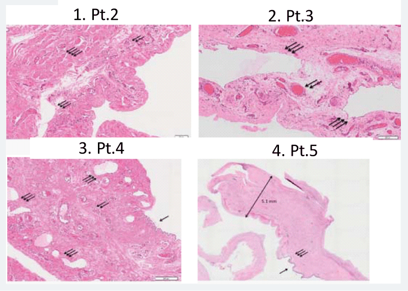
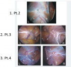
Figure 4.4:
Representative histopathological findings of the hydrocele wall (H&E original magnification x 100). 1 Pt.2 (Y.K.), Thickened hydrocele wall containing dilated lymph vessels and cremaster muscle fibers. 2 Pt.3 (N.S.), Proliferation of markedly dilated lymph vessels. 3 Pt.4 (N.F.), A wall lined by flattened mesotherial cells, containing proliferated blood vessels and lymph vessels. Neutrophil infiltrations are scattered. 4 Pt.5 (Y.I.), Loupe magnification showing a thickened tunica vaginalis wall, reaching up to 5.1 mm in thickness, with dense connective tissue. Most of mesothelial linings are degenerated and falling. Abbreviations: arrow, mesothelial linings; double arrow, blood vessels; triple arrow, lymph vessels; fourfold arrow, cremaster muscle fibers.
Read Full Article HTML DOI: 10.29328/journal.ascr.1001015 Cite this Article Read Full Article PDF
More Images
Similar Articles
-
Laparoscopic Adrenalectomy; A Short Summary with Review of LiteratureMushtaq Chalkoo*,Naseer Awan,Hilal Makhdoomi,Syed Shakeeb Arsalan ,Waseem Raja. Laparoscopic Adrenalectomy; A Short Summary with Review of Literature . . 2017 doi: 10.29328/journal.ascr.1001001; 1: 001-011
-
Bouveret Syndrome in an Elderly FemaleZvi H. Perry*, Udit Gibor,Shahar Atias,Solly Mizrahi,Alex Rosental, Boris Kirshtein. Bouveret Syndrome in an Elderly Female . . 2017 doi: 10.29328/journal.ascr.1001002; 1: 012-015
-
Intestinal obstruction complicated by large Morgagni herniaMartín Arnau B*,Medrano Caviedes R,Rofin Serra S, Caballero Mestres F,Trias Folch M. Intestinal obstruction complicated by large Morgagni hernia . . 2017 doi: 10.29328/journal.ascr.1001003; 1: 016-020
-
Clinical significance of Urinary Amylase in Acute PancreatitisMumtaz Din Wani,Mushtaq Chalkoo*,Zaheer Ahmed,Awhad Mueed Yousuf,Yassar Arafat,Syed Shakeeb Arsalan,Jaffar Hussain. Clinical significance of Urinary Amylase in Acute Pancreatitis . . 2017 doi: 10.29328/journal.ascr.1001004; 1: 021-031
-
Use of Orthodeoxia by pulse Oximetry in the detection of Hepatopulmonary SyndromeCesar Raul Aguilar Garcia*,Guadalupe Viridiana Ontiveros Guerra. Use of Orthodeoxia by pulse Oximetry in the detection of Hepatopulmonary Syndrome . . 2017 doi: 10.29328/journal.ascr.1001005; 1: 038-041
-
Surgery and new Pharmacological strategy in some atherosclerotic chronic and acute conditionsLuisetto M*,Nili-Ahmadabadi B,Ghulam Rasool Mashori. Surgery and new Pharmacological strategy in some atherosclerotic chronic and acute conditions . . 2017 doi: 10.29328/journal.ascr.1001006; 1: 042-048
-
The revolution of cardiac surgery evolution Running head: Cardiac surgery evolutionMarzia Cottini*. The revolution of cardiac surgery evolution Running head: Cardiac surgery evolution . . 2017 doi: 10.29328/journal.ascr.1001007; 1: 049-050
-
Dieulafoy’s Lesion related massive Intraoperative Gastrointestinal Bleeding during single Anastomosis Gastric Bypass necessitating total Gastrectomy: A Case ReportAshraf Imam,Khalayleh Harbi*,Miller Rafael, Khoury Deeb,Buyeviz Victor,Guy Pines,Sapojnikov Shimon. Dieulafoy’s Lesion related massive Intraoperative Gastrointestinal Bleeding during single Anastomosis Gastric Bypass necessitating total Gastrectomy: A Case Report . . 2017 doi: 10.29328/journal.ascr.1001008; 1: 051-055
-
Laparoscopic partial nephrectomy-does tumor profile influence the operative performance?Krishanu Das*, George P Abraham, Kishnamohan Ramaswai, Datson George P,Jisha J Abraham,Thomas Thachill, Oppukeril S Thampan. Laparoscopic partial nephrectomy-does tumor profile influence the operative performance? . . 2017 doi: 10.29328/journal.ascr.1001009; 1: 056-060
-
Comments for the Nuremberg Code 70 Years LaterJie Zhang,Chao-Jun Kong, Zhong Jia*. Comments for the Nuremberg Code 70 Years Later . . 2017 doi: 10.29328/journal.ascr.1001010; 1: 061-061
Recently Viewed
-
Reverse Breech Extraction versus Vaginal Push before Uterine Incision during Cesarean Section with Fully Dilated Cervix and Impacted Fetal HeadElsayed Elshamy*, Abdelbar Sharaf, Abdelhamid Shaheen. Reverse Breech Extraction versus Vaginal Push before Uterine Incision during Cesarean Section with Fully Dilated Cervix and Impacted Fetal Head. Clin J Obstet Gynecol. 2023: doi: 10.29328/journal.cjog.1001145; 6: 160-164
-
Age as a Predictor of Time to Response for Patients Undergoing Medical Management of Endometrial CancerM Larissa Weirich*, Carolyn R Larkins, Wendy Y Craig, Emily Meserve, Terri Febbraro, Jason Lachance, Leslie S Bradford. Age as a Predictor of Time to Response for Patients Undergoing Medical Management of Endometrial Cancer. Clin J Obstet Gynecol. 2023: doi: 10.29328/journal.cjog.1001144; 6: 150-159
-
A Comparative Study of Metoprolol and Amlodipine on Mortality, Disability and Complication in Acute StrokeJayantee Kalita*,Dhiraj Kumar,Nagendra B Gutti,Sandeep K Gupta,Anadi Mishra,Vivek Singh. A Comparative Study of Metoprolol and Amlodipine on Mortality, Disability and Complication in Acute Stroke. J Neurosci Neurol Disord. 2025: doi: 10.29328/journal.jnnd.1001108; 9: 039-045
-
Exploring the Potential of Medicinal Plants in Bone Marrow Regeneration and Hematopoietic Stem Cell TherapyUgwu Okechukwu Paul-Chima*,Alum Esther Ugo. Exploring the Potential of Medicinal Plants in Bone Marrow Regeneration and Hematopoietic Stem Cell Therapy. Int J Bone Marrow Res. 2025: doi: 10.29328/journal.ijbmr.1001019; 8: 001-005
-
Satellite-Based Analysis of Air Pollution Trends in Khartoum before and After the ConflictHossam Aldeen Anwer*,Abubakr Hassan,Ghofran Anwer. Satellite-Based Analysis of Air Pollution Trends in Khartoum before and After the Conflict. Ann Civil Environ Eng. 2025: doi: 10.29328/journal.acee.1001074; 9: 001-011
Most Viewed
-
Evaluation of Biostimulants Based on Recovered Protein Hydrolysates from Animal By-products as Plant Growth EnhancersH Pérez-Aguilar*, M Lacruz-Asaro, F Arán-Ais. Evaluation of Biostimulants Based on Recovered Protein Hydrolysates from Animal By-products as Plant Growth Enhancers. J Plant Sci Phytopathol. 2023 doi: 10.29328/journal.jpsp.1001104; 7: 042-047
-
Sinonasal Myxoma Extending into the Orbit in a 4-Year Old: A Case PresentationJulian A Purrinos*, Ramzi Younis. Sinonasal Myxoma Extending into the Orbit in a 4-Year Old: A Case Presentation. Arch Case Rep. 2024 doi: 10.29328/journal.acr.1001099; 8: 075-077
-
Feasibility study of magnetic sensing for detecting single-neuron action potentialsDenis Tonini,Kai Wu,Renata Saha,Jian-Ping Wang*. Feasibility study of magnetic sensing for detecting single-neuron action potentials. Ann Biomed Sci Eng. 2022 doi: 10.29328/journal.abse.1001018; 6: 019-029
-
Pediatric Dysgerminoma: Unveiling a Rare Ovarian TumorFaten Limaiem*, Khalil Saffar, Ahmed Halouani. Pediatric Dysgerminoma: Unveiling a Rare Ovarian Tumor. Arch Case Rep. 2024 doi: 10.29328/journal.acr.1001087; 8: 010-013
-
Physical activity can change the physiological and psychological circumstances during COVID-19 pandemic: A narrative reviewKhashayar Maroufi*. Physical activity can change the physiological and psychological circumstances during COVID-19 pandemic: A narrative review. J Sports Med Ther. 2021 doi: 10.29328/journal.jsmt.1001051; 6: 001-007

HSPI: We're glad you're here. Please click "create a new Query" if you are a new visitor to our website and need further information from us.
If you are already a member of our network and need to keep track of any developments regarding a question you have already submitted, click "take me to my Query."


