Figure 2
Carotid artery disease: AngioCT features
Joana Ferreira*, Olinda Miranda, Alexandre Lima Carneiro, Sandrina Braga, João Correia Simões, Celso Carrilho, Amílcar Mesquita and Jorge Cotter
Published: 26 July, 2019 | Volume 3 - Issue 2 | Pages: 059-060

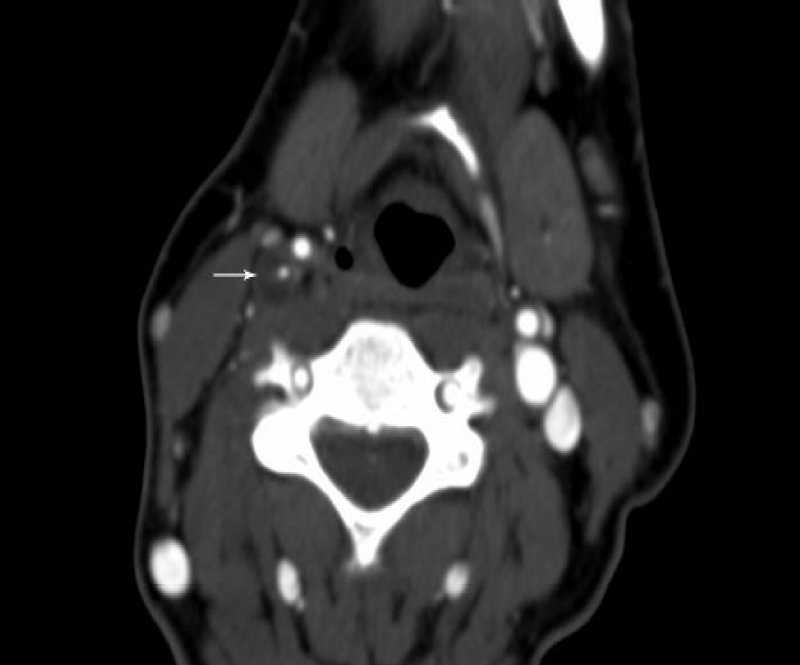
Figure 2:
Coronal TC image of the right internal carotid artery hematoma. In this image is clear that the carotid lumen is surrounded by a hypo dense mural thickening, from the hematoma (arrow). The hematoma between the intima and media and causes the vessel diameter expansion. The enhancing vessel wall is visible due to contrast enhancement of vasa vasorum in the adventitial layer.
Read Full Article HTML DOI: 10.29328/journal.ascr.1001035 Cite this Article Read Full Article PDF
More Images
Similar Articles
-
Gossypiboma due to a retained surgical sponge following abdominal hysterectomy, complicated by intestinal migration and small bowel obstruction- A Case ReportVivek Agrawal,Praroop Gupta*. Gossypiboma due to a retained surgical sponge following abdominal hysterectomy, complicated by intestinal migration and small bowel obstruction- A Case Report. . 2018 doi: 10.29328/journal.ascr.1001017; 2: 015-017
-
Laparoscopic Cholecystectomy: Challenges faced by beginners our perspectiveKunal Chowdhary,Gurinder Kaur,Kapil Sindhu,Muzzafar Zaman*,Aliya Shah,Rohit Dang,Ashish Kumar,Jose John Maiakal,Ashutosh Bawa. Laparoscopic Cholecystectomy: Challenges faced by beginners our perspective. . 2018 doi: 10.29328/journal.ascr.1001018; 2: 018-024
-
Carotid artery disease: AngioCT featuresJoana Ferreira*,Olinda Miranda,Alexandre Lima Carneiro,Sandrina Braga,João Correia Simões,Celso Carrilho, Amílcar Mesquita,Jorge Cotter. Carotid artery disease: AngioCT features. . 2019 doi: 10.29328/journal.ascr.1001035; 3: 059-060
-
Beginnings of bariatric and metabolic surgery in SpainAniceto Baltasar*. Beginnings of bariatric and metabolic surgery in Spain. . 2019 doi: 10.29328/journal.ascr.1001042; 3: 082-090
-
Stomach cancer: epidemiological, diagnostic and therapeutic aspects at the Kara Teaching Hospital, TogoDOSSOUVI Tamegnon*,EL-HADJI YAKOUBOU Rafiou,ADABRA Komlan,AMAVI Ayi,AMOUZOU Efoé-Ga Olivier,KANASSOUA Kokou Kouliwa,KASSEGNE Iroukora,DOSSEH Ekoué David. Stomach cancer: epidemiological, diagnostic and therapeutic aspects at the Kara Teaching Hospital, Togo. . 2022 doi: 10.29328/journal.ascr.1001062; 6: 001-003
-
CVS: An Effective Strategy to Prevent Bile Duct InjurySardar Rezaul Islam*, Debabrata Paul, Shah Alam Sarkar, Mohammad Hanif Emon, Tania Ahmed. CVS: An Effective Strategy to Prevent Bile Duct Injury. . 2024 doi: 10.29328/journal.ascr.1001080; 8: 027-031
Recently Viewed
-
Crime Scene Examination of Murder CaseSubhash Chandra*,Pradeep KR,Jitendra P Kait,SK Gupta,Deepa Verma. Crime Scene Examination of Murder Case. J Forensic Sci Res. 2024: doi: 10.29328/journal.jfsr.1001071; 8: 108-110
-
Associations of Burnout, Secondary Traumatic Stress and Individual Differences among Correctional Psychologists`Irina G. Malkina-Pykh*. Associations of Burnout, Secondary Traumatic Stress and Individual Differences among Correctional Psychologists`. J Forensic Sci Res. 2017: doi: 10.29328/journal.jfsr.1001003; 1: 018-034
-
An overview of the influence of climate change on food security and human healthSanober Naheed*. An overview of the influence of climate change on food security and human health. Arch Food Nutr Sci. 2023: doi: 10.29328/journal.afns.1001044; 7: 001-011
-
Evaluating the Pros and Cons of Evening and Weekend Outpatient Medical Imaging: Implications for Patients and Radiology ProfessionalsLucas Cohen and Ethan Cohen*. Evaluating the Pros and Cons of Evening and Weekend Outpatient Medical Imaging: Implications for Patients and Radiology Professionals. J Radiol Oncol. 2024: doi: 10.29328/journal.jro.1001069; 8: 078-084
-
Radiation SafetyBradley Anderson,Greg Anderson*. Radiation Safety. J Radiol Oncol. 2024: doi: 10.29328/journal.jro.1001072; 8: 097-099
Most Viewed
-
Feasibility study of magnetic sensing for detecting single-neuron action potentialsDenis Tonini,Kai Wu,Renata Saha,Jian-Ping Wang*. Feasibility study of magnetic sensing for detecting single-neuron action potentials. Ann Biomed Sci Eng. 2022 doi: 10.29328/journal.abse.1001018; 6: 019-029
-
Evaluation of In vitro and Ex vivo Models for Studying the Effectiveness of Vaginal Drug Systems in Controlling Microbe Infections: A Systematic ReviewMohammad Hossein Karami*, Majid Abdouss*, Mandana Karami. Evaluation of In vitro and Ex vivo Models for Studying the Effectiveness of Vaginal Drug Systems in Controlling Microbe Infections: A Systematic Review. Clin J Obstet Gynecol. 2023 doi: 10.29328/journal.cjog.1001151; 6: 201-215
-
Prospective Coronavirus Liver Effects: Available KnowledgeAvishek Mandal*. Prospective Coronavirus Liver Effects: Available Knowledge. Ann Clin Gastroenterol Hepatol. 2023 doi: 10.29328/journal.acgh.1001039; 7: 001-010
-
Causal Link between Human Blood Metabolites and Asthma: An Investigation Using Mendelian RandomizationYong-Qing Zhu, Xiao-Yan Meng, Jing-Hua Yang*. Causal Link between Human Blood Metabolites and Asthma: An Investigation Using Mendelian Randomization. Arch Asthma Allergy Immunol. 2023 doi: 10.29328/journal.aaai.1001032; 7: 012-022
-
An algorithm to safely manage oral food challenge in an office-based setting for children with multiple food allergiesNathalie Cottel,Aïcha Dieme,Véronique Orcel,Yannick Chantran,Mélisande Bourgoin-Heck,Jocelyne Just. An algorithm to safely manage oral food challenge in an office-based setting for children with multiple food allergies. Arch Asthma Allergy Immunol. 2021 doi: 10.29328/journal.aaai.1001027; 5: 030-037

HSPI: We're glad you're here. Please click "create a new Query" if you are a new visitor to our website and need further information from us.
If you are already a member of our network and need to keep track of any developments regarding a question you have already submitted, click "take me to my Query."


