Figure 1
Extremely large hemangioma of the liver: Safety of the expectant management
Subhi Mansour, Sharbel Khouri, Maisa Andraous and Safi Khuri*
Published: 06 September, 2019 | Volume 3 - Issue 2 | Pages: 061-064
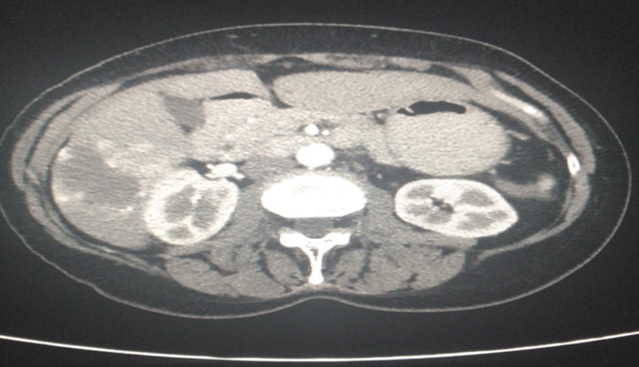
Figure 1:
An axial abdominopelvic Computed Tomography (CT) scan demonstrates an incidental finding of giant hepatic hemangioma at the right lobe of the liver. CT scan was done due to suspicion of diverticulitis of the sigmoid colon.
Read Full Article HTML DOI: 10.29328/journal.ascr.1001036 Cite this Article Read Full Article PDF
More Images
Similar Articles
-
Extremely large hemangioma of the liver: Safety of the expectant managementSubhi Mansour, Sharbel Khouri,Maisa Andraous,Safi Khuri*. Extremely large hemangioma of the liver: Safety of the expectant management. . 2019 doi: 10.29328/journal.ascr.1001036; 3: 061-064
Recently Viewed
-
Investigation of Stain Patterns from Diverse Blood Samples on Various SurfacesSonia Rajkumari*. Investigation of Stain Patterns from Diverse Blood Samples on Various Surfaces. J Forensic Sci Res. 2024: doi: 10.29328/journal.jfsr.1001061; 8: 028034
-
Management of Non-contact Injuries, Nonspecific Chronic Pain, and Prevention via Sensory Conflicts Detection: Vertical Heterophoria as a Landmark IndicatorEric Matheron*. Management of Non-contact Injuries, Nonspecific Chronic Pain, and Prevention via Sensory Conflicts Detection: Vertical Heterophoria as a Landmark Indicator. J Nov Physiother Rehabil. 2024: doi: 10.29328/journal.jnpr.1001057; 8: 005-013
-
Multipurpose Antioxidants based on Food Industry Waste: Production and Properties EvaluationToshkhodjaev*. Multipurpose Antioxidants based on Food Industry Waste: Production and Properties Evaluation. Arch Food Nutr Sci. 2025: doi: 10.29328/journal.afns.1001062; 9: 001-003
-
The Role of Buspirone in the Treatment of Patients with Paranoid Schizophrenia with Non-Suicidal Auto Aggression Who Have Had a Coronavirus InfectionIV Kravchenko*. The Role of Buspirone in the Treatment of Patients with Paranoid Schizophrenia with Non-Suicidal Auto Aggression Who Have Had a Coronavirus Infection. Arch Psychiatr Ment Health. 2023: doi: 10.29328/journal.apmh.1001047; 7: 010-014
-
Flood Risk Management in South-west Nigeria: Lagos as a Case StudyIdowu Michael*. Flood Risk Management in South-west Nigeria: Lagos as a Case Study. Ann Civil Environ Eng. 2024: doi: 10.29328/journal.acee.1001073; 8: 096-097
Most Viewed
-
Feasibility study of magnetic sensing for detecting single-neuron action potentialsDenis Tonini,Kai Wu,Renata Saha,Jian-Ping Wang*. Feasibility study of magnetic sensing for detecting single-neuron action potentials. Ann Biomed Sci Eng. 2022 doi: 10.29328/journal.abse.1001018; 6: 019-029
-
Evaluation of In vitro and Ex vivo Models for Studying the Effectiveness of Vaginal Drug Systems in Controlling Microbe Infections: A Systematic ReviewMohammad Hossein Karami*, Majid Abdouss*, Mandana Karami. Evaluation of In vitro and Ex vivo Models for Studying the Effectiveness of Vaginal Drug Systems in Controlling Microbe Infections: A Systematic Review. Clin J Obstet Gynecol. 2023 doi: 10.29328/journal.cjog.1001151; 6: 201-215
-
Prospective Coronavirus Liver Effects: Available KnowledgeAvishek Mandal*. Prospective Coronavirus Liver Effects: Available Knowledge. Ann Clin Gastroenterol Hepatol. 2023 doi: 10.29328/journal.acgh.1001039; 7: 001-010
-
Causal Link between Human Blood Metabolites and Asthma: An Investigation Using Mendelian RandomizationYong-Qing Zhu, Xiao-Yan Meng, Jing-Hua Yang*. Causal Link between Human Blood Metabolites and Asthma: An Investigation Using Mendelian Randomization. Arch Asthma Allergy Immunol. 2023 doi: 10.29328/journal.aaai.1001032; 7: 012-022
-
An algorithm to safely manage oral food challenge in an office-based setting for children with multiple food allergiesNathalie Cottel,Aïcha Dieme,Véronique Orcel,Yannick Chantran,Mélisande Bourgoin-Heck,Jocelyne Just. An algorithm to safely manage oral food challenge in an office-based setting for children with multiple food allergies. Arch Asthma Allergy Immunol. 2021 doi: 10.29328/journal.aaai.1001027; 5: 030-037

HSPI: We're glad you're here. Please click "create a new Query" if you are a new visitor to our website and need further information from us.
If you are already a member of our network and need to keep track of any developments regarding a question you have already submitted, click "take me to my Query."


