More Info
Author's satisfaction with
- Friendly and hassle-free publication process
- Less production time of articles
- Constructive peer-review
- Enhancing journal reputation
- Regular feedback system
- Quick response to authors' queries
Recently Viewed
Most Viewed
Clinical Images
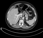
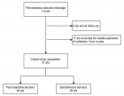
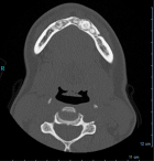
Table of Contents

Laparoscopic approach for acute right iliac fossa pathology: Our experience
Published on: 31st December, 2020
OCLC Number/Unique Identifier: 8882678494
Laparoscopic approach in emergency theatre is an irreplaceable tool to manage patients with acute surgical pathology. We retrospectively reviewed surgical access records from the Emergency Department for acute right iliac fossa pathology. We considered 51 patients (16 male, 35 female, mean age 23.8 years) access for acute right iliac fossa pathology over the last year. 44 patients underwent laparoscopic approach (86%); 8 patients were treated with an open approach. Outcomes evaluation was based on data comparison from open appendicectomy over 4 year time period.
Variables considered for data analyses were: role of laparoscopic surgery for gangrenous/perforated appendicitis, Conversion rate, Laparoscopy appendicectomy for elderly patients.
Our study demonstrated that a laparoscopic approach at acute right iliac fossa pathology is feasible, safe and can offer a low incidence of infectious complications, less post-operative pain, rapid recovery, and represent a valid diagnostic tool in doubtful cases, at the expense of longer operating time than OA. We suggest that LA should be the initial choice for all patients with acute right iliac fossa pathology.
Acute abdomen as complication of a knee arthroscopy: A case report
Published on: 22nd December, 2020
OCLC Number/Unique Identifier: 8872656452
A knee arthroscopy in spinal anaesthesia was performed on a 67 years old male patient. During the procedure the patient was hemodynamically stable, until he suddenly turned pale and started complaining of severe pain in lower abdomen with signs of guarding. The procedure was finished as urgently as possible and after releasing the tourniquet we noticed significant difference in volume of the leg, with redness distal to tourniquet. Urgent lab results were essentially unremarkable and the patient was sent for the urgent radiological diagnostics.
CD of the left leg described fluid in the soft tissues of the thigh, scrotum, and abdomen; and the unenhanced CT of the abdomen showed free fluid along the entire femoral shaft of the left thigh, extending towards pelvis and abdomen to perihepatic and perisplenic space, and retroperitoneum, with gas bubbles tracking along anterior aspect of the left thigh into the left retroperitoneum.
He was admitted to the ICU, and within few hours all symptoms have resolved and his further recovery was without complications.
Intentıonal reımplantatıon of hopeless teeth: Cases serıes
Published on: 31st October, 2020
OCLC Number/Unique Identifier: 8872657975
Intentional replantation is an alternative for the treatment of advanced periodontal destruction of the anterior teeth. Systemically healthy three female patient was referred to our clinic with functional complaints. Diagnosis were chronic periodontitis and class III mobility was noted at the mandibular incisors with complete periodontal attachment loss. After phase I periodontal treatment periodontally involved teeth were extracted, endodontic treatment accomplished, the teeth were replanted and fixed to its place with fiber reinforced composite splint. At the end of 2 years tooth was in function with alveolar bone gain. Intentional replantation provided long-term maintenance of patient’s own teeth.
The role of robotic segmentectomy for non-small cell lung cancer
Published on: 6th October, 2020
OCLC Number/Unique Identifier: 8682303248
Segmentectomy may be applied to all segments; superior segmentectomies (lower lobe superior segments for both lungs), lingulectomies (two segments forming lingulas of upper left lobe) and basal segmentectomies (segments other than superior segment for both lungs). In lung segment resections; segmentectomy has an equivalent morbidity, recurrence and survival rate compared to lobectomy, in patients with stage I lung cancer, tumors smaller than 2 cm and within the segmental anatomical limits. Segmentectomy also contributes more to preserving lung function and exercise capacity than lobectomy. In tumor resection; especially in patients with advanced age, insufficient performance or insufficient cardiopulmonary reserve, 2 cm in diameter and acceptable segmental margins may be provided.
Limited long-term results show oncological results of robotic approach similar to open and VATS approaches. Robotic approach facilitates surgery with more intuitive movements, greater flexibility and high definition, three-dimensional vision. However, high cost and lack of touch sense are main disadvantages of robotic surgery. New studies are needed to assess quality of life, morbidity, oncological results and cost effectiveness. However, considering development of technology in our age and fact that many surgical robot brands will be released in the near future, it is predicted that disadvantages of robotic surgery will be minimized in the near future.
This article reviews experience of segmentectomy in non-small cell lung cancer and discusses benefits and limitations of robotic segmentectomy.
Video-assisted thoracic surgery in advanced non-small cell lung cancer treatment
Published on: 14th August, 2020
OCLC Number/Unique Identifier: 8653118841
Non-small cell lung cancer is one of the leading causes of cancer-related mortality worldwide. Despite recent advances in adjuvant treatments, surgical resection is basis of treatment. With the development of minimally invasive surgery in thoracic surgery, surgeons work on minimally invasive surgery for advanced stages of lung cancer, previously considered non-operable at all or previously considered non-operable with minimally invasive surgery approach.
Minimally invasive surgical techniques which are routinely used in the surgical treatment of early-stage lung cancer have started to be treated in more complicated and advanced stages of lung cancer. Bilateral anatomic resections, operations after neoadjuvant chemotherapy, bronchial sleeve lobectomies, double sleeve lobectomies, complementary pneumonectomies, and carinal sleeve resections can be performed by minimally invasive methods. The option of video-assisted surgery should be considered with oncological principles at foreground if patients have acceptable lung and cardiac performance conditions with minimal comorbidities.
This study reviews VATS experience in patients with advanced-stage lung cancer worldwide and discusses potential benefits and limitations of using VATS technology to perform thoracic surgery procedures.
Anticipation of difficulty during laparoscopic cholecystectomy
Published on: 24th July, 2020
OCLC Number/Unique Identifier: 8639116784
Background: Laparoscopic cholecystectomy (LC), is one of the most commonly performed surgical procedures worldwide, it is accepted as the gold standard in the treatment of symptomatic gallstones for its minimal invasiveness, less pain and early recovery.
Purpose: To predict the difficulty of laparoscopic cholecystectomy in patients according to the recently published scoring system and select the difficult cases to be done by a senior surgeon.
Patients: This is a prospective cohort study. This study took place Oct 6th University Hospital and Kasr El Aini Hospital, Cairo university; the study involved 120 patients admitted with calcular cholecystitis, arranged for laparoscopic cholecystectomy.
Methods: Laparoscopic cholecystectomy after applying the scoring system.
Results: In our study we found that age, sex and ultrasonographic data were significant predictive factors for assessment preoperatively difficult cases that will be operated upon. We found 14 patients above 50 years who scored to be difficult and very difficult were at outcome difficult, only three patients converted to open surgery over fifty.
Conclusion: We can report that obese patient who were over fifty with history of previous upper abdominal surgery and ultrasonographic picture showed thick walled GB and pericholecystic collection had high risk of conversion. At this study scoring system was used for prediction of difficult laparoscopic cholecystectomy sensitivity was 93.75% and specificity was 52.94% of the scoring system at score 5 for prediction of easy or difficult laparoscopic cholecystectomy.
Leakage after sleeve gastrectomy: Endoscopic stenting VS surgical intervention
Published on: 24th July, 2020
OCLC Number/Unique Identifier: 8639114108
Background: Laparoscopic sleeve gastrectomy (LSG) is becoming more popular in the treatment of obesity. LSG is safe with a low morbidity. The complications rarely result in morbidity and even mortality. Leaks are the major complication associated with LSG with a reported prevalence between 1.9% and 2.4%.
Objective: To compare surgical intervention and endoscopic stenting for treatment of gastric leakage after sleeve gastrectomy.
Patients and method: Our study included 30 patients presented with post sleeve leaks discovered by routine postoperative imaging or during the follow up period. Patients were recruited from October 6th university hospital during the period from August 2017 to August 2019. Patients were divided to the following groups: 1) Endoscopy group: This included 15 patients with post sleeve leakage undergoing endoscopic stent insertion. 2) Surgery group: which included 15 patients with post sleeve leak age undergoing surgical management. This division was random.
Results: Our study showed that Endoscopic stenting for management of post sleeve gastrectomy leakage is an effective method with lower morbidity and shorter post-operative hospital stay than surgical management. Some patients may be good candidates for early surgical intervention in type 1 leakage if managed early before dissemination of leakage and before tissues become friable. Complications of stents include stent migration (26%), stent related ulcer (13%) and stricture (13%). while the surgical intervention carries more complications (DVT, chest infection, wound infection and stricture) and longer postoperative hospital stay.
Conclusion: endoscopic management of post-sleeve gastrectomy leakage with stenting is recommended because it successfully manages the leaks and avoids invasive procedures with less risk, with shorter hospital stay and early return of function.
When conservative treatment in trachea laserations?
Published on: 21st July, 2020
OCLC Number/Unique Identifier: 8639105303
Introduction: The tracheobronchial injuries are usually fatal and some of the lucky people can reach emergency services without dying in the place of trauma. They can cause severe symptoms which can be lifetreathing. This type of injuries must been taken carefully and need to decide fast what treatment you going to give.
Case report: We present a 53 years old patient who has been stabbed during a fight and got his trachea ruptured. His complaints shortness of breath and neck swelling. He can be treated conservatively with bronchoscopic and clinical evaluation.
Discussion: Tracheobronchial injuries are life-threatening and the airway must be secured first. They can be treated conservatively in some cases. CT can be useful but fiberoptic bronchoscopy is the key in diagnosis.
Conclusion: Although early treatment of tracheal lacerations is urgent surgery, it is reported that these injuries can be treated with conservative methods under appropriate conditions.
Acute necrotising pancreatitis masquerading as psoas abscess: A report of two cases
Published on: 15th July, 2020
Acute pancreatitis is commonly diagnosed clinically, with its classical presentation of upper abdominal pain, backed by raised serum levels of enzymes amylase and lipase. However, unusual presentation of this common surgical emergency as a psoas abscess is a rare finding which can lead to missed diagnosis with a fatal outcome.
We present here two such cases of acute necrotising pancreatitis masquerading as psoas abscess, with no classical clinical symptoms and only mildly raised levels of serum amylase and lipase. The region of pancreas involved by necrosis influenced the site of presentation of the psoas abscess. In the first case, acute necrotising pancreatitis involving head and neck of pancreas presented as psoas abscess presenting in the right lumbar region, while the left side collection due to pancreatitis involving body and tail of pancreas manifested as an abscess in left flank.
While evaluating the aetiology of a psoas abscess, a differential diagnosis of necrotizing pancreatitis should be kept as a possibility.
Atypical Ductal Hyperplasia: Factors predicting upstaging to carcinoma
Published on: 6th July, 2020
OCLC Number/Unique Identifier: 8633817372
Aim: Percutaneous core needle biopsy (CNB) is considered the gold standard technique for initial histological diagnosis of suspicious breast lesions seen on screening mammogram, but it is less reliable for diagnosing atypical ductal hyperplasia (ADH) due to significant rates of diagnosis upstaging to malignant disease after excision biopsy. The purpose of this study was to identify factors that predict diagnosis upstage to carcinoma in patients diagnosed with ADH on core biopsy.
Methods: A retrospective database search identified 52 consecutive CNB of suspicious breast lesions revealing pure ADH. Inclusion criteria included asymptomatic women presenting for screening mammogram, who subsequently underwent surgical excision. Logistic regression analysis evaluated clinical, radiological, and histological factors.
Results: A total of 52 patients with ADH on CNB were identified who met our criteria. Twenty-six of 52 patients (50%) were upstaged to ductal carcinoma in situ or invasive carcinoma, based on histological interpretation of the surgically excised specimen. Lesion size was showed to be a statistically significant predictor on univariable logistic regression analysis. Multivariate logistic regression analysis revealed Asian ethnicity and lesion size as independent predictors of malignancy (p = 0.050 and 0.011, respectively). Conversely, women of Middle Eastern and European origin and lesions < 15 mm on mammography were negative predictors of malignancy.
Conclusion: Lesion size ≥ 15 mm on mammography and Asian ethnicity are independent risk factors for breast carcinoma in asymptomatic patients diagnosed with ADH on CNB.
Contact
Select by Volume & Issue
Most Viewed Keywords
- COVID-19
- His bundle
- Ischemic heart disease
- Forensic science
- Techno-econophysics
- Ca2+/cAMP interaction
- Childbirth
- Intracerebral hemorrhage
- Pancreatic fistula
- COVID-19
- Toumbak
- Elderly
- Citrus
- Glomus tumors, rare tumor, glomus body
- Mat Activities
- Bilateral sagittal split osteotomy
- Neuromyelitis
- PET TAC
- Electromyography
- Gamma flow detector
University/Institution
Select and search by University/Institution.
Articles by Country
Select and search by country to get related articles.


Testmonials

I would like to thank JPRA for taking this decision. I understand the effort it represents for you. I'm truly happy to have the paper published in JPRA. And I'll certainly consider JPRA for my next publications as I was satisfied of the service provided, the efficiency and promptness of the interactions we had.
Emmanuel BUSATO
Publishing with the International Journal of Clinical and Experimental Ophthalmology was a rewarding experience as review process was thorough and brisk. Their visibility online is second to none as their published articles appear in all search engines. I will encourage researchers to publish with them.
Elizabeth Awoyesuku
“The choice to submit the forensic case study to the Journal of Addiction Therapy and Research was dictated by the match between the content and the potential readership. The publication process proved to be expedient and we were provided with constructive feedback from reviewers. The final article layout is attractive and conforms to standards. All-in-all, it has been a rewarding process.”
Elisabeth H Wiig
Archives of Vascular Medicine is one of the top class journal for vascular medicine with highly interesting topics. You did a professional and great Job!
Elias Noory
Thank you very much. I think the review process and all of what concerns the administration of the publication concerning our paper has been excellent. The nice and quick answers have been very good I think.
Doris Nilsson
Journal of Pulmonary and Respiratory Research is good journal for respiratory research purposes. It takes 2-3 weeks maximum for review of the manuscript to get published and any corrections to be made in the manuscript. It needs good articles and studies to get publish in the respiratory medicine. I am really glad that this journal editors helped me to get my case report published.
Divya Khanduja
Thanks you and your colleague for the great help for our publication. You always provide prompt responses and high quality of service. I am so happy to have you working with me. Thanks again!
Diana (Ding) Dai
Service and process were excellent as was the “look” of the article when published.
Deane Waldman
Great, thank you! It was very efficient working w/ your group. Very thorough reviews (i.e., plagiarism, peer, etc.). Would certainly recommend that future authors consider working w/ your group.
David W Brett
Your services are very good
Chukwuka Ireju Onyinye
I very much appreciate the humanitarian services provided in my stead by this journal/publisher. It exhibits total absence of editorial impertinence. As an Author, I have been guided to have a fruitful experience. The editorial care is highly commendable.
Chrysanthus Chukwuma
"An amazing experience with the Journal of Advanced Pediatrics and Child Health. Very fast blind review with pertinent corrections and suggestions. I highly recommand both the journal and the editor."
Chaimae Khairoun
The submission is very easy and the time from submission to response from the reviewers is short. Correspondence with the journal is nice and rapid.
Catrin Henriksson
The Clinical Journal of Obstetrics and Gynecology is an open access journal focused on scientific knowledge publication with emphasis laid on the fields of Gynecology and Obstetrics. Their services toward us have been encouraging through their kindness and respect. Great consideration has been given to us as young budding researchers and we are very grateful for this.
Carole Assontsa
During the process your positive communication, prompt feedback and professional approach is very highly appreciated. We would like to thank you very much for your support.
Can Vuran
I do appreciate for your service including submission, analysis, review, editorial and publishing process. I believe these esteemed journal enlighten the science with its high-quality personel.
Bora Uysal
I am very much pleased with the fast track publication by your reputed journal's editorial team. It is really helpful for researchers like me from developing nations. I strongly recommend your journal for publication.
Badri Kumar Gupta
It has been a fabulous journey writing articles for your journal because of the encouragement you people provide for writers from developing nations like India. Kindly continue the same. Looking forward for a long term association.
Badareesh Lakshminarayana
Many thanks for publishing my article in your great journal and the friendly and hassle-free publication process, the constructive peer-review, the regular feedback system, and the Quick response to any queries.
Azab Elsayed Azab
I would like to thank this journal for publishing my Research Article. Something I really appreciate about this journal is, they did not take much time from the day of Submission to the publishing date. Looking forward to have more publications in future.
Ayush Chandra
Submission of paper was smooth, the review process was fast. I had excellent communication and on time response from the editor.
Ayokunle Dada
Your service is very good and fast reply, also your service understand our situation and support us to publication our articles.
Ayman M Abu Mustafa
Really good service with prompt response. Looking forward to having long lasting relationship with your journal
Avishek Bagchi
Your service is excellent. Processing and editing were very fast. I hope to publish more of my works in your journal.
Ausraful Islam
I wanna to thank Clinical Journal of Nursing Care and Practice for its effort to review and publish my manuscript. This is reputable journal. Thank you!
Atsedemariam Andualem
“It was a delightful experience publishing my manuscript with the Clinical Journal of Obstetrics and Gynecology. They offered me lots of opportunities I never had from most publishing houses and their prompt services are greatly appreciated.”
Asafo Jones
I hope to ability to make some new investigation and publish in Your Company in future.
Artur Stopyra
I like the quality of the print & overall service. The paper looks quite impressive. Hope this will attract interested readers. All of you have our best wishes for continued success.
Arshad Khan
Your big support from researchers around the world is the best appreciation from your scientific teams. We believe that there should be no barrier in science and you make it real and this motto come true.
Arefhosseinir Rafi
Your journal co-operation is very appreciable and motivational. I am really thankful to your journal and team members for the motivation and collaboration to publish my work.
Assistant Professor, UCLAS Uttaranchal University, Dehradun, India
Archna Dhasmana
I am glad to submit the article to Heighten Science Publications as it has a very smooth and fast peer-review process, which enables the researchers to communicate their work on time.
Anupam M
This is to specify that I have had an extensive and detailed interaction with the Editorial team of Annals of Clinical Gastroenterology and Hepatology, USA, lasting over a significant period of time. My interaction has been extremely pleasant, especially with Ms Allie Smith, as I find the communication quite inspiring and crystal clear. The attitude of aforesaid individuals is quite helpful and guiding in pertinent instances. It has been a commemorative journey so far working with the Journal and I hope that the symbiosis will continue, evolve and flourish in the forthcoming years. I wish the journal, related personnel and aforementioned individuals a fruitful, successful run.
New Delhi, India
Anubha Bajaj
We appreciate the fact that you decided to give us full waiver for the applicable charges and approve the final version. You did an excellent job preparing the PDF version. Of course we will consider your magazine for our future submissions and we will pay the applicable fees then.
Anna Dionysopoulou
''Co-operation of Archives of Surgery and Clinical Research journal is appreciable. I'm impressed at the promptness of the publishing staff and the professionalism displayed. Thank you very much for your support, help and encouragement.''
Anıl Gokce
Congratulations for the excellence of your journal and high quality of its publications.
Angel MARTIN CASTELLANOS
The service from the journal staff has been excellent.
Andy Smith
I was very pleased with the quick editorial process. We are sure that our paper will have great visibility, among other things due to its open access. We believe in science accessible to all.
Anderson Fernando de Souza
It was a great experience publishing through JCICM. The article has reached out to several institutions. Appreciate your professional work. Hope to work with you again
Anas Wardeh
Publishing an article is a long process, but working with your publication department made things go smoothly, even though the process took exactly 5 months from the time of submitting the article till the time I have favourable response, the missing part is the peer review details, which is essential in self auditing and future improvement, overall experience was excellent giving your understanding of the situation of lack of financial institution support.
Anas Diab
I think that Heighpubs very good. You are very helpful. Thank you for everything.
Ana Ribeiro
Regarding to be services, we note that are work with high standards of professionalism translated into quick response, efficiency which makes communication accessible. Furthermore, I believe to be much inviting for the submission of future works for publication purposes.
Amélia João Alice Nkutxi
I would like to mention that I had a wonderful experience working with HSPI. The whole process right from manuscript submission to peer review till the publication of the article was very prompt & efficient. I wish you good luck for the future.
Amarjeet Gambhir
Once I submitted the manuscript, the response time of the reviewers was very fast. The fine-tuning of the galley proof was likewise prompt. I believe the journal provide a valuable outlet to disseminate physical rehabilitation scientific knowledge to the clinical community. Respectfully. Dr. Alon
Alon
We really appreciate and thanks the full waiver you provide for our article. We happy to publish our paper in your journal. Thank you very much for your good support and services.
Ali Abusafia
It was a real pleasure working with your team. The review was done fast, and it was very clear, the editing was flawless, the article was published quickly compared to other journals, and everyone was understanding and helpful. I will gladly recommend this journal to my acquaintances in academia.
Alexandra Cozma
To the editorial team at HSPI and the Journal of Clinical Nephrology: Thank you so much for your hard work and collaboration in bringing our article to life. Your staff was responsive, flexible, and communicative and made the process smooth and easy. Thank you!
Alejandro Munoz
Dear colleagues! I am satisfied with our cooperation with you. Your service is at a high level. I hope for a future relationship. Let me know if I can get a paper version of the magazine with my articles from you. I see them on the Internet.
Aksenov V.V
"This is my first time publishing with the journal/publisher. I am impressed at the promptness of the publishing staff and the professionalism displayed. Thank you for encouraging young researchers like me!"
Ajite Kayode
I want to thank you for our collaboration. You were fast and effective with a positive spirit of teamwork. I am truly excited from our collaboration. You were like always fast, efficient and accurate. I hope that in the near future we will collaborate again.
Aikaterini Solomou
In my opinion, you provide a very fast and practical service.
Ahmet Eroglu
Great, We are too comfortable with the process including the peer review process and quality. But, the journal should be indexed in different databases such scopus.
Afework Edmealem
We really appreciate your efforts towards our article, the professional way you handle our request for exemption from charges. It was a great honor for us to publish in your magazine.
Achraf elbakkaly
I really liked the ease of submitting my manuscript in the HSPI journal. Further, the peer review was timely completed and I was communicated the final decision on my manuscript within 10 days of submission which is really appreciable. I strongly recommend all the scientists and researchers to submit their work in this journal”
Abu Bashar
My candid opinion is that the service you render is second to none. My favourite part is the prompt response to issue, really i value that.
Abiodun Akanbi Adeogun
Thank you very much for accepting our manuscript in your journal “International Journal of Clinical Virology”. We are very thankful to the esteemed team for timely response and quick review process. The editorial team of International Journal of Clinical Virology is too cooperative and well-mannered during the publication process. We are hopeful to publish many quality papers in your journal and I suggest the International Journal of Clinical Virology to all of my colleagues, researchers and friends to publish their research here.
Abdul Baset
I, Muhammad Sarwar Khan, am serving as Editor on Archives of Biotechnology and Biomedicine (ABB). I submitted an editorial titled, 'Edible vaccines to combat Infectious Bursal Disease of poultry' for publication in ABB. After submitting the manuscript; the services rendered by the management and technical personnel to handle and process the manuscript were marvelous. Plagiarism report was shared with me with complements before reviewers' comments, All steps including article processing and service charges were well taken care of keeping in view the author's interest/preference. All together, it was an encouraging and wonderful experience working with ABB personnel.
University of Agriculture, Pakistan
Muhammad Sarwar Khan
Your journal has accomplished its intended mission of providing very effective and efficient goals in dealing with submissions, conducting the reviewing process and in publishing accepted manuscripts in a timely manner. Keep up the great work and services that you provide.
University of Jacqmar, Inc., USA
John St. Cyr
I am to express my view that Heighten Science Publications are reliable quick even after peer review process. I hope and wish the publications will go a long way in disseminating science to many interested in scientific research.
College of Fisheries, CAU(I), Tripura, India
Ajit Kumar Roy
The Journal Clinical Nephrology provides a good opportunity for readers to stay updated in the field of clinical nephrology. Additionally - it provides a good opportunity for authors to publish their work. 1. Publication of the accepted manuscripts is sufficiently rapid. 2. The trust factor between the journal and me, as an author, is very important and well preserved. 3. Peer review process very rapid and effective.
Assaf Harofeh Medical Center, Israel
Leonid Feldman
In 2017, I submitted a manuscript to the journal Archives of Biotechnology and Biomedicine belonging to Heighten Science Publications Corporation. Within one week I already received the response from the editor. All processing steps were really fast so in terms of a speedy publication I can particularly recommend the journal Archives of Biotechnology and Biomedicine. The responsible contact person of the journal was always available, which gives a trustworthy impression to the author. Also the peer review process was clear and constructive. So from my experience with Heighten Science Publications Corporation I can recommend publishing there.
University of Tubingen, Germany
Yvonne Mast
We thank to the heighten science family, who speed up the publication of our article and provide every support.
Mehmet Besir
The services of the journal were excellent. The most important thing for an author is the speed of the peer review which was really fast here. They returned in a few days and immediately replied all of my questions. I want to refer this platform to all scholars. Many thanks.
Eastern Mediterranean University, Cyprus
Zehra Guchan TOPCU
Thank you for your attitude and support. I am sincerely grateful to you and the entire staff of the magazine for the high professionalism and fast quality work. Thank you very much!
USA
Igor Klepikov
Thank you and your company for effective support of authors which are very much dependable on the funds gambling for science in the different countries of our huge and unpredictable world. We are doing our work and should rely on a teams like Galley Proof-HSPC. Great success to all of you for the 2019th! Be well all the year long.
Russia
Victor V Apollonov
The editorial process was quickly done. The galley proof was sent within a week after being accepted for publication. The editorial team was very helpful and responded promptly.
India
Rohit Kulshrestha
Publishing with the International Journal of Clinical and Experimental Ophthalmology was a rewarding experience as review process was thorough and brisk. Their visibility online is second to none as their published articles appear in all search engines. I will encourage researchers to publish with them.
University of Port Harcourt Teaching Hospital, Nigeria
Dr. Elizabeth A Awoyesuku
"It was a pleasure to work with the editorial team of the journal on the submission of the manuscript. The team was professional, fast, and to the point".
NC A&T State University, USA
Moran Sciamama-Saghiv
Submission of paper was smooth, the review process was fast. I had excellent communication and on time response from the editor.
Ekiti State University Teaching Hospital, Nigeria
Ayokunle Dada
I am delighted and satisfied with. Heighten Science Publications as my manuscript was thoroughly assessed and published on time without delay. Keep up the good work.
Ido-Ekiti/Afe Babalola University, Nigeria
Dr. Shuaib Kayode Aremu
"This is my first time publishing with the journal/publisher. I am impressed at the promptness of the publishing staff and the professionalism displayed. Thank you for encouraging young researchers like me!"
Ekiti State University, Nigeria
Adebukola Ajite
I wanna to thank clinical journal of nursing care and practice for its effort to review and publish my manuscript. This is reputable journal. Thank you!
Wollo University, Ethiopia
Atsedemariam Andualem
We appreciate your approach to scholars and will encourage you to collaborate with your organization, which includes interesting and different medical journals. With the best wishes of success, creativity and joy in life, prosperity in the medical field.
Ivano- Frankivsk National Medical University, Ukraine
Nataliya Kitsera
Thank you very much for your support and encouragement. I am truly impressed by your tolerance and support. Thank you very much
Diaverum: PADC, Jeddah, Saudi Arabia
Nasrulla Abutaleb
You are such a nice person. Your journal co-operation is very appreciable and motivational.
Department of Biotechnology, Uttaranchal college of Applied and Life Sciences, Uttaranchal University, Dehradun, Uttarakhand, India
Archna Dhasmana
“Mobile apps and wearable technology are becoming ubiquitous in our environment. Their integration with healthcare delivery is just beginning to take shape. The early results are promising and the possibilities great."
BS, PharmD., MBA, CPHIMS, FHIMSS, Adjunct Professor, Global Healthcare Management, MCPHS University, Chief Strategy Offi cer, MedicaSoft, Senior Advisor, National Health IT (NHIT) Collaborative for Underserved, New York HIMSS, National Liaison, Health 2.0 Boston, Past Chair, Chair Innovation, USA
Helen Figge
“The choice to submit the forensic case study to the Journal of Addiction Therapy and Research was dictated by the match between the content and the potential readership. The publication process proved to be expedient and we were provided with constructive feedback from reviewers. The final article layout is attractive and conforms to standards. All-in-all, it has been a rewarding process.”
Ph.D, Boston University Department of Communication Sciences and Disorders and Knowledge Research Institute, Inc., 2131 Reflection Bay Drive, Arlington, Texas 76013, USA
Elisabeth H. Wiig
The service is nice and the time of processing the application is fast.
Department of Neurosurgery, Queen Elizabeth Hospital, Hong Kong
Long Ching
Your service is very good and fast reply, Also your service understand our situation and support us to publication our articles.
Palestine College of Nursing, Khan Younis, Gaza Strip, Palestine
Ayman M Abu Mustafa
“It was a delightful experience publishing my manuscript with the Clinical Journal of Obstetrics and Gynecology. They offered me lots of opportunities I never had from most publishing houses and their prompt services are greatly appreciated.”
Department of Agricultural Economics, Agribusiness and Extension, Kwame Nkrumah University of Science and Technology, Kumasi, Ghana
Akowuah Jones Asafo
Related Journals

HSPI: We're glad you're here. Please click "create a new Query" if you are a new visitor to our website and need further information from us.
If you are already a member of our network and need to keep track of any developments regarding a question you have already submitted, click "take me to my Query."